
Some enzymes, such as proteases like trypsin, rely solely on amino acid chemistry and water to facilitate catalysis. Other enzymes need a helping hand, in the form of metal ions such as magnesium or iron, while many others deploy a limited range of low molecular weight species, collectively termed coenzymes, or cofactors.
The biological universality of the cofactors adenosine di- and triphosphate (AD/TP) and nicotinamide adenine dinucleotide (NAD), for example), suggests an early appearance on the evolutionary landscape. The marshalling of metals by the enzymes present in emerging life forms is perhaps much easier to accept, since they were and remain relatively abundant on Earth, and can be imported by a dedicated protein; much less energy demanding that a dedicated, multi-step, biosynthetic pathway.
Before discussing examples of cofactor-dependent biocatalysis, let me point out the considerable scale differences between the naturally hexameric, NAD-dependent glutamate dehydrogenase from the anaerobe Clostridium symbiosum, (shown left) and its six, bound cofactors (NAD): the amino acid substrate glutamate is oxidatively deaminated by the enzyme in a key, metabolic reaction.
Let me begin by clarifying the difference between some cofactors and substrates (or co-substrates) and introduce some of the language used by enzymologists. I shall use cofactor as a blanket term in this post; but there are some important distinctions. The term co-substrate is applied to those cofactors, such as ATP, that can act as a second substrate, for example in donating a phosphate group to a glucose substrate at the active site of hexokinase, to generate gluse-6-phosphate and ADP. Similarly, NADH (the reduced form of NAD+), in the presence of ammonia ions, reduces 2-oxo-glutarate (also called alpha ketoglutarate) to glutamate and in the process, NADH is oxidised. Cofactors are sometimes referred to as co-enzymes and are usually bound with a very high affinity (with dissociation constants in the 10-1000 nM range). Sometimes cofactors are covalently bound to the enzyme: these are often referred to as prosthetic groups. In addition, an enzyme when isolated in complex with its coenzyme is often referred to as a holo-enzyme. When the enzyme is free from its coenzyme, it is called an apo-enzyme. Enzymes that rely on metal ions which include many protein-based enzymes and most of the known, RNA based enzymes (typically, Mg2+ ions form structural and catalytic cofactors), are referred to as metallo-enzymes. In some cases, catalytic RNA molecules are called ribozymes: however, some authors collectively refer to protein and RNA based enzymes as biocatalysts.
In any given cell, at any time, the number of different types of substrate molecules far exceeds the number of cofactor molecules. In other words, the Venn diagram for enzymes and cofactors looks quite different than that for enzymes and substrates. The amino acid sequence determinants specifying the stereo-chemical recognition and engagement between enzymes and cofactors is characterised by lower information entropy than that between enzymes and their substrates.
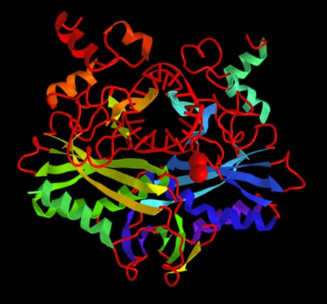
The enzyme shown on the left is a restriction enzyme (more precisely the Type II restriction endonuclease EcoRV). These enzymes were pivotal in the early stages of the molecular cloning revolution from the 1970s onwards. They recognise and cleave a specific sequence of nucleotides in the “middle” of a double stranded segment of DNA, which may be chemically synthesised or a biological sample of genomic DNA, for example. (The DNA double helix is shown in red running down its long axis through the active site of the enzyme in the figure on the right). These enzymes deserve a whole post in themselves, but I shall use them here as an example of metallo-enzymes, where the two red spheres indicate the location of two magnesium cations (often replaced in crystallography by calcium or manganese for experimental reasons). In the absence of magnesium, the enzyme will not cleave the DNA. The metal ions do not just help, without them there is no catalysis observed. Similarly, the addition of magnesium ions to DNA in solution, has no effect on the integrity of the DNA. Clearly, a mutual interdependency exists between cofactor and enzyme, a situation that is almost universal among restriction enzymes with the notable exception of BfiI. The precise role of the magnesium ions in each restriction enzymes is likely to be different from enzyme to enzyme, but in general terms, without these metal ions, the critical positioning of active site amino acids in the stabilisation of the transition state, is not achieved.
A special case of metallo-enzymes are ribozymes. I remember in the mid-’80s poring over the papers from two independent labs: Tom Cech at Boulder, Colorado, and Sidney Altman’s lab at Yale, claiming that they had demonstrated catalytic activity of RNA, in the absence of proteins. I can only imagine what the atmosphere was like in their lab meetings: both scientists had great track records and would have been ultra-sceptical of any data from their graduate students and post-docs, especially when they were about to overturn the established view that all enzymes are proteins. The rest is history, and a share in the 1989 Nobel Prize for Chemistry by Cech and Altman! In retrospect, one of the milestones in protein synthesis was the determination of the structure of the first transfer RNA (tRNA), which resembles something of a cross between a small protein and a double helical nucleic acid. Maybe this was hinting at the potential for RNA molecules to behave like proteins? The problem facing a nucleic acid in folding into a globular structure (common amongst protein enzymes) is the electrostatic challenge of its negatively charged primary structure. By adsorbing cations, such as magnesium, the repulsive forces from the negatively charged polynucleotide are neutralised, and the RNA is able to adopt a stable, compact structure. In this way, metal ions are acting as a structural cofactor, and in addition, they contribute to the stabilisation of the transition state, in a manner similar to that observed in restriction endonucleases. In Nature, the small but significant number of ribozymes that have been biochemically analysed (take a look at Dan Herschlag’s lab homepage at Stanford for more information) have enhanced our understanding of the principles of biocatalysis.
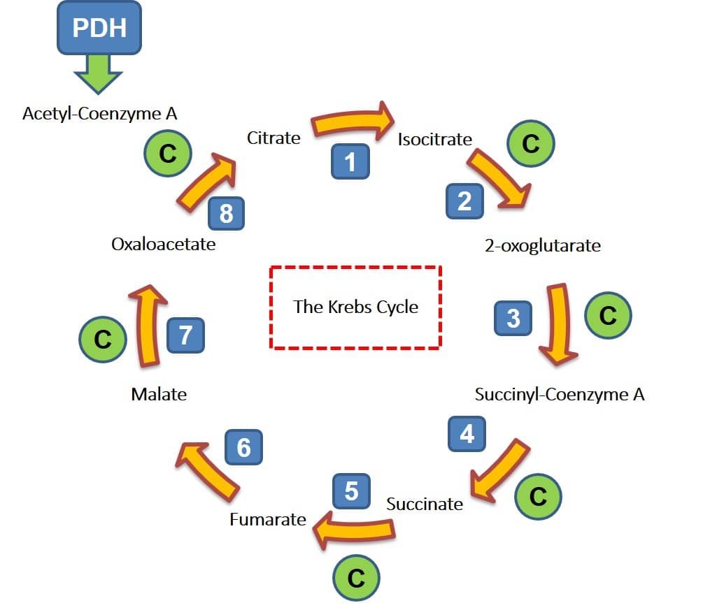
All biochemists are aware of the fact that some biochemical reactions are more challenging to catalyse than others. The challenge may be physical: the substrates may be sparingly soluble in water or the target bonds are poorly accessibly. Alternatively, the thermodynamic barriers to product formation may be too high. There are other reasons, but these are amongst the most common. The Krebs Cycle (shown left) lies at the heart of oxidative metabolism across all aerobic organisms from microbes to plants and animals. Through a sequence of eight, enzyme catalysed reactions, every mol of acetyl Co-enzyme A (AcCoA) (which may be derived from glycolysis via pyruvate, or fatty acid oxidation), combines with oxaloacetate to generate citrate, which is then transformed with the concomitant release of 3 mol NADH, one mol FADH and 2 mol GTP. The process yields a modest amount of energy in the form of GTP, but it is the energy derived indirectly via NADH and FADH via the electron transfer chain, that powers aerobic cells. Where there is involvement of a cofactor, I have indicated this with a letter C in a green circle, while the enzyme reactions are numbered. It is the roles of NADH and FADH that I am most interested in here. I’ll focus on the enzyme 2-oxoglutarate dehydrogenase (step 3) and succinate dehydrogenase, the enzyme at step 5. For those interested in a more in depth perspective on the biological significance of the Krebs Cycle, I can recommend Transformer (by Nick Lane, 2022).
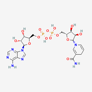
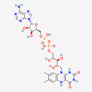
The two molecules shown above are nicotinamide adenine dinucleotide (NAD) and flavin adenine dinucleotide (FAD). In terms of rigour, I should point to the formal nomenclature for these two cofactors. The oxidised form of NAD is written as NAD+, to reflect the positive charge on the nicotinamide ring nitrogen (bottom right hand side). In its reduced state, it is generally written as NADH. In the case of FAD, there are two reduced forms, FADH and FADH2, the isoalloxazine ring also at the lower right hand side of the structure, can accept 2 separate additions of 1 H+ and 1 e− to form the half- and subsequently fully reduced forms. The aromatic nature of the isoalloxazine ring is lost upon reduction, thereby raising its energetic state. The redox state of FAD, gives rise to characteristic spectral features, which are often perturbed during catalysis and sometimes simply by interactions in different enzyme binding sites. NADH shows a significant absorption at 340nm, making redox reactions easy to follow using a conventional spectrophotometer. In fact, spectral “visibility” of NAD and FAD dependent enzymes (as well as haem containing proteins), made them the focus of many enzymologists during the 20th century.
Both cofactors are characterised by the presence of an adenine ring, ribose sugars and phosphates, a feature you will recognise with respect to the energy currency of most living cells, ATP and of course the nucleic acid RNA. Common threads of evolution. It is perhaps not too surprising that enzymes utilising these cofactors share some common primary and tertiary structural motifs, and much of this occupied the minds of structural biologists and protein sequencers before the breakthroughs in molecular cloning in the 1980s opened the way for structural biologists and gene sequencers to explore enzymes more widely. NAD and FAD are cofactors that participate in the reaction pathway of enzymes, if they facilitate the reduction of a substrate, they are then themselves reduced (and vice versa). As a result, cofactor recycling mechanisms exist and before a second catalytic event occurs, a cofactor must be returned to its “active” form. By comparison, a metal ion cofactor is more likely to facilitate the polarisation of amino acid side chains, active site water molecules, or the incoming substrate, and therefore do not require recycling in the same way.
The 2-oxo acid dehydrogenase reaction (interchangeably called alpha-ketoglutarate dehydrogenase) in the Krebs Cycle (number 3 in the cycle diagram), can be written in its simplest form as:
The reaction is an oxidative decarboxylation and the role of NAD is part of a much more complex sequence of reactions (for those interested, these enzymes are multi-protein complexes, on a size scale similar to ribosomes, and the detailed roles of each of the components is very well documented, the perspective by one of the pioneers of the field, Lester Reed is available here, and is open access). The main point I wish to draw to your attention is that cofactors of the NAD class, are active participants in catalysis, and are often considered as substrates, especially in kinetic analyses. However, at any one time in a cell, there are many more substrates than cofactors, which in my view places cofactors like NAD and FAD, as something in between a substrate and a catalyst. Moreover, cofactors like FAD and NAD share structural groups that facilitate docking into their partner enzymes, suggesting common genetic origins for cofactor recognition.
In the case of the succinate dehydrogenase reaction, in which succinate is converted to fumarate, the cofactor is FAD and similarly, the participation of the cofactor in the reaction requires complex regeneration. In contrast, enzymes like alcohol and lactate dehydrogenases, mainstays of 20th century enzymology, derive their NAD/NADH from a cellular pool. In the case of FAD- dependent enzymes, the cofactor is usually much more firmly bound to the enzyme, and in some cases is covalently linked to the protein chain. As you will have no doubt realised by now, nucleoside cofactors like NAD and FAD have been co-opted by enzymes in several different ways to support catalysis, but in all cases, they provide the fundamental redox support chemistry for all life forms.
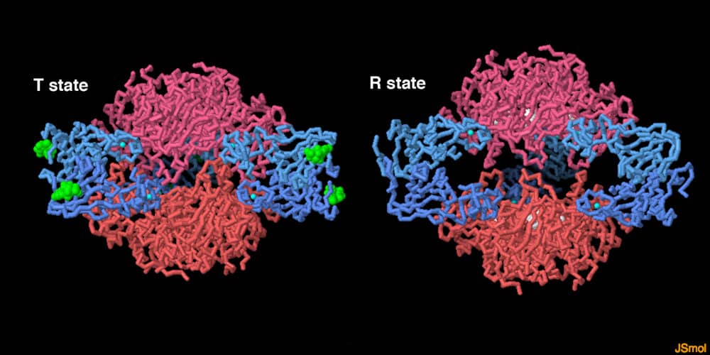
The beautiful image above shows the three dimensional structure of the multi-subunit (12 in all) enzyme Aspartate Transcarbamoylase (ATCase) that catalyses the first committed step in pyrimidine biosynthesis. I have chosen this enzyme partly for sentimental reasons: firstly, it was one of the topics in my undergraduate degree that fascinated me, but more importantly it is a superb example of an allosterically regulated enzyme. The word allostery (a combination of “other” and “site”) was first coined by Jacob and Monod, pioneers in the field of enzyme regulation, both genetically and kinetically. For me, allostery is both an elegant and apposite term, which matches the remarkable intellectual contributions made by these two French polymaths during the 1950s and ’60s in particular. The labels R (relaxed) and T (tense) refer to two stable equilibrium conformational states promoted by the binding of an allosteric ligand. I am going to include allosteric effectors in my group of molecules referred to as cofactors: these are ligands (ranging from small metabolites to proteins) that induce conformational transitions upon binding to a specific protein. In the case of ATCase, the final product of the pyrimidine biosynthesis pathway, CTP, when bound to a regulatory subunit of the enzyme, decreases its catalytic velocity. In contrast, the binding of ATP, a purine, increases catalytic velocity. In this way the cell exerts remarkably fine control over the proportions of the purines and pyrimidines synthesised, which form a key part of DNA, RNA and the cofactors discussed above. From an enzyme prospecting perspective, failure to add a positive allosteric effector may limit the detection of an enzyme activity. And of course by way of contrast, the presence of a negative allosteric effector may suppress the enzyme activity you may be looking for. In short, small molecules such as ATP, can significantly stimulate (positive allostery) or inhibit (negative allostery) the activity of certain enzymes. Such enzymes are often said to exhibit cooperativity: initially this was assumed to be limited to positive cooperativity, where the addition of the allosteric ligand stimulates reaction velocity. The theoretical existence of negative cooperativity, although less common has also been observed.
In an attempt to bring the discussion to a close, I hope I have alerted you to the importance of cofactors in understanding both the power and limitations of enzymes in catalysis. Understanding the sequence of events that led to the emergence of the first living organisms on Earth, remains a topic for research, but it is clear that a limited set of cofactors have come to pervade all forms of life. These include NAD, FAD, A(D)TP and a small group of metal ions. In attempting to detect a new enzyme activity, or modifying the properties of an enzyme, it is advisable to consider the potential role that may be played by cofactors. In addition, when searching for enzymes in complex cell extracts, an essential cofactor for your enzyme, may be a good substrate for another. These challenges can be overcome by regularly supplementing assay mixtures with potential cofactors, or even designing a cofactor regeneration system as part of your activity screening protocol. Finally, this post is meant to provide a taster for appreciating the properties of cofactors, understanding cofactor dependency in a specific class of enzyme activity will require a deeper evaluation of that enzyme.
